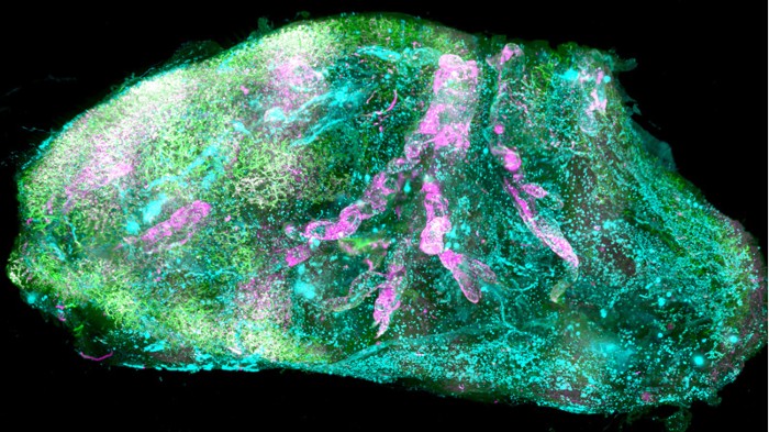
Human organs made transparent reveal their inner secrets
Date: 14.2.2020
Machine learning helps to expose the molecules and structures within a human kidney, eye and thyroid.
 To study the body’s inner workings, scientists generally chop up organs and construct 3D images from several thin slices of tissue – a time-consuming and error-prone process. Now, researchers have developed a way to peer inside intact human organs in microscopic detail.
Ali Ertürk at the Munich Helmholtz Centre in Germany and his colleagues soaked organs in chemicals that preserve the structure of tissues while stripping them of fats and pigments that normally block the passage of light. The process makes tissues permeable to dyes and molecules that label specific structures, such as neurons and blood vessels.
To take 3D images of the clear organs, the researchers used a microscopy technique that helped them to image thin slices of tissue without having to cut into it. Then, the team developed machine-learning algorithms to rapidly analyse millions of individual cells in the images.
Using this approach, the researchers took detailed snapshots of an intact human eye, thyroid and kidney. The method could help to reveal human organ functions in health and disease, the researchers say.


