Worlds smallest imaging device can 3D-scan inside your blood vessels
Date: 22.7.2020
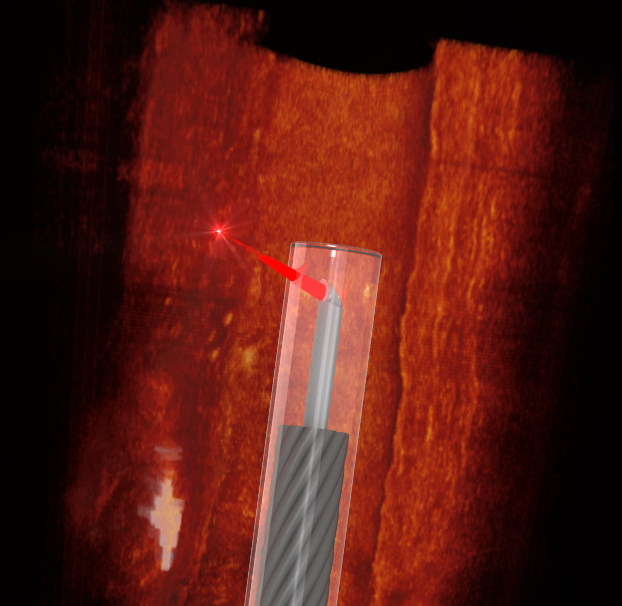
An Australian/German team has developed the world's smallest imaging device, at the thickness of a human hair.
 It's capable of traveling down the blood vessels of mice, offering unprecedented abilities to 3D-scan the body at microscopic resolutions.
To build this miniature endoscope, the team took a fine optical fiber with a diameter of less than half a millimeter (0.02 in), including its protective sheath. The researchers then used a 3D micro-printing technique to print a minuscule side-facing lens into it with a diameter less than 0.13 mm (0.005 in) – too small to be seen with the naked eye.
This optical fiber was then connected to an optical coherence tomography (OCT) scanner as a flexible probe. OCT is a 3D depth-sensitive scanning technology, commonly used to map the retina in optometry and ophthalmology. It uses near-infrared light to penetrate into tissue, measuring the wave interference between a reference beam and a probe beam to build up live 3D images that can look through surfaces within the body into the structures beneath at microscopic resolutions.
The team performed successful tests of the device in both human and mouse blood vessels, demonstrating its ability to deliver quality OCT images and the flexibility to get where it needs to go in the body.


